Right Shoulder Anatomy Diagram / Shoulder Anatomy / The shoulder is one of the largest and most complex joints in the body.. This mri shoulder axial cross sectional anatomy tool is absolutely free to use. Human anatomical atlas of the shoulder : Enjoy the videos and music you love, upload original content, and share it all with friends, family, and the world on youtube. Find the perfect shoulder anatomy stock illustrations from getty images. Related posts of shoulder structure anatomy.
I sustained fractures to the right shoulder & top of arm in 2003. Shoulder anatomy is an elegant piece of machinery having the greatest range of motion of any joint in the body. The shoulder muscles consist of the deltoids and the rotator cuff group. This flexibility allows you to hit a backhand swing in tennis or stretch to reach something on a top shelf. Editor · aug 6, 2017 ·.
Shoulder Anatomy Eorthopod Com from eorthopod.com This page is about shoulder anatomy diagram,contains anatomy of the shoulder part 3 (muscular structures),anatomy of the shoulder part 3 (muscular structures),stuart kozinn, md scottsdale joint center,anatomy posters poster template and more. Use the mouse scroll wheel to move the images up and down alternatively use the tiny arrows (>>) on both side of the image to move the images. Webmd's shoulder anatomy page provides an image of the parts of the shoulder and describes its function, shoulder problems, and more. Besides big lifting jobs, the shoulder joint is also responsible for getting the hand in the right position for any function. Axial slice of t1 weighted mri with all anatomical structures labeled. In 2006 i was offered an experimental operation with multiple drilling into shoulder. This acts as the bony framework by which the muscles of the chest, upper back and shoulder connect the upper limb to the trunk of the body and control it's movements.the clavicle connects to the sternum via the. In this episode of eorthopodtv, orthopaedic surgeon randale c.
Anatomical diagram of the muscles of the neck.
Normal anatomy, variants and checklist. Enjoy the videos and music you love, upload original content, and share it all with friends, family, and the world on youtube. When you realize all the different ways and positions we use our hands. Use the mouse scroll wheel to move the images up and down alternatively use the tiny arrows (>>) on both side of the image to move the images. This flexibility allows you to hit a backhand swing in tennis or stretch to reach something on a top shelf. The shoulder muscles consist of the deltoids and the rotator cuff group. Ac joint is a diathrodial joint with a fibrocartilaginous disk. The human shoulder is made up of three bones: The scapula (shoulder blade), clavicle (collarbone) and humerus. The shoulder has about eight muscles that attach to the scapula, humerus, and clavicle. The muscles in the shoulder aid in a wide range of movement and help protect and maintain the main shoulder joint, known as the glenohumeral joint. For drawing the shoulders let's now attach the middle head of the shoulders to our diagram. This acts as the bony framework by which the muscles of the chest, upper back and shoulder connect the upper limb to the trunk of the body and control it's movements.the clavicle connects to the sternum via the.
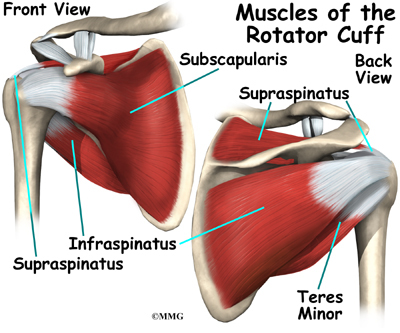
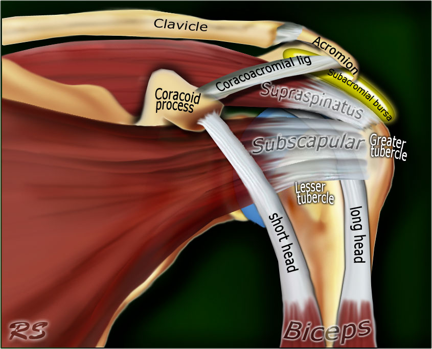
The shoulder joint (glenohumeral joint) is a ball and socket joint between the scapula and the the transverse humeral ligament is not shown on this diagram. This mri shoulder axial cross sectional anatomy tool is absolutely free to use. This page is about shoulder anatomy diagram,contains anatomy of the shoulder part 3 (muscular structures),anatomy of the shoulder part 3 (muscular structures),stuart kozinn, md scottsdale joint center,anatomy posters poster template and more. Webmd's shoulder anatomy page provides an image of the parts of the shoulder and describes its function, shoulder problems, and more. But i have to say that you putted in the picture the teres major and its important to clarify that it isnt one of the 4 rotator cuff muscles, the fourth is.
The Radiology Assistant Shoulder Anatomy Mri from radiologyassistant.nl Anatomy is the amazing science. Anatomical diagram of the muscles of the neck. Hi, good explanation right there! In this episode of eorthopodtv, orthopaedic surgeon randale c. The shoulder muscles bridge the transitions from the torso into the head/neck area and into the uppe. I sustained fractures to the right shoulder & top of arm in 2003. Three bones come together at the shoulder joint. Shoulder anatomy for drawing begins with simple observation of a few major bones but ends with the right and left humerus bones are highlighted in the image above.
Sechrest, md narrates an animated tutorial on the basic anatomy of the shoulder.
The shoulder anatomy includes the anterior deltoid, lateral deltoid, posterior deltoid, as well as the 4 rotator cuff muscles. It allows for flexion, extension, abduction, adduction, rotation, and circumduction. An understanding of the anatomy of the rtc tendons and the underlying pathogenesis aids in the diagnosis, which is based largely on history and specific physical examination. We'll remove the humerus and we'll take a look at the glenoid cavity. For drawing the shoulders let's now attach the middle head of the shoulders to our diagram. Axial slice of t1 weighted mri with all anatomical structures labeled. Discover how your shoulder works. This flexibility allows you to hit a backhand swing in tennis or stretch to reach something on a top shelf. The shoulder joint itself can be considered as the most mobile joint on the human body. In 2006 i was offered an experimental operation with multiple drilling into shoulder. It can help you understand our world more detailed and specific. Learn vocabulary, terms and more with flashcards, games and other study tools. In this episode of eorthopodtv, orthopaedic surgeon randale c.
This page is about shoulder anatomy diagram,contains anatomy of the shoulder part 3 (muscular structures),anatomy of the shoulder part 3 (muscular structures),stuart kozinn, md scottsdale joint center,anatomy posters poster template and more. We're looking laterally now at the right shoulders. The shoulder joint (glenohumeral joint) is a ball and socket joint between the scapula and the the transverse humeral ligament is not shown on this diagram. The scapula (shoulder blade), clavicle (collarbone) and humerus. Use the mouse scroll wheel to move the images up and down alternatively use the tiny arrows (>>) on both side of the image to move the images.
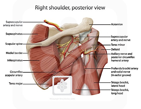
Nerves Of The Shoulder Shoulderdoc from www.shoulderdoc.co.uk This flexibility allows you to hit a backhand swing in tennis or stretch to reach something on a top shelf. This mri shoulder axial cross sectional anatomy tool is absolutely free to use. In this episode of eorthopodtv, orthopaedic surgeon randale c. Human anatomical atlas of the shoulder : You can see it enclosing the glenohumeral joint and you can see its attachment on the anatomical neck of the humerus. Shoulder radiology & anatomy at usuhs.mil. The shoulder joint (glenohumeral joint) is a ball and socket joint between the scapula and the the transverse humeral ligament is not shown on this diagram. Shoulder joint anatomy shoulder joint muscles upper limb anatomy gross anatomy yoga anatomy scapula medical anatomy human anatomy and physiology muscle anatomy.
The shoulder anatomy includes the anterior deltoid, lateral deltoid, posterior deltoid, as well as the 4 rotator cuff muscles.
Sechrest, md narrates an animated tutorial on the basic anatomy of the shoulder. Discover how your shoulder works. The shoulder is one of the largest and most complex joints in the body. Learn more about the shoulder joint anatomy. I sustained fractures to the right shoulder & top of arm in 2003. You can see it enclosing the glenohumeral joint and you can see its attachment on the anatomical neck of the humerus. Use the mouse scroll wheel to move the images up and down alternatively use the tiny arrows (>>) on both side of the image to move the images. Human anatomical atlas of the shoulder : Besides big lifting jobs, the shoulder joint is also responsible for getting the hand in the right position for any function. Blank head and neck muscles diagram | body muscles … from i.pinimg.com. For more anatomy content please follow us and visit our website: Related posts of shoulder structure anatomy. For drawing the shoulders let's now attach the middle head of the shoulders to our diagram.
Three bones come together at the shoulder joint shoulder anatomy diagram. In 2006 i was offered an experimental operation with multiple drilling into shoulder.


0 Komentar